Hip And Upper Thigh Anatomy - Human Anatomy and Physiology of Muscles Online on | Human ... / This deep muscle begins in the low back and pelvis and connects on the inside edge of the upper femur.
Hip And Upper Thigh Anatomy - Human Anatomy and Physiology of Muscles Online on | Human ... / This deep muscle begins in the low back and pelvis and connects on the inside edge of the upper femur.. Want to learn more about it? 2, tensor fasciae latae m. Muscles of hip and thigh: Anatomynote.com found upper thigh muscle anatomy … related posts the anatomical areas found on the upper limb can serve as key landmarks to help us find important anatomical structures such as finding one of the. Its quadrangular shape and flat design allow it to adduct and flex the hip joint.
340 anatomical structures of the hip region were labeled, accessible on anatomical parts: Superficial layer right side, anterior view. The femoral artery is a continuation of the. In order to help understand the conditions causing hip pain and their surgical treatment, it is important to first have a basic understanding of the anatomy of the hip and how it functions. The uppermost of the medial thigh muscles is the pectineus muscle.
Muscles of the Posterior Thigh - Hamstrings - Damage ... from teachmeanatomy.info Anatomynote.com found upper thigh muscle anatomy … related posts the anatomical areas found on the upper limb can serve as key landmarks to help us find important anatomical structures such as finding one of the. 2, tensor fasciae latae m. Along the upper portion of the thigh, just lateral to the gracilis, the adductor longus muscle is ranked as the most anterior of this group of thigh muscles. The adductor muscle on the inner thigh; Longest muscle in the body. The information contained in anatomy atlases is not a substitute for the medical care and advice of your physician. Upper limb anatomy arm anatomy muscle anatomy anatomy study body anatomy anatomy thigh: Superficial layer right side, anterior view.
Hip flexor deep in pelvis a composite o… used to extend the hip when climbing st…
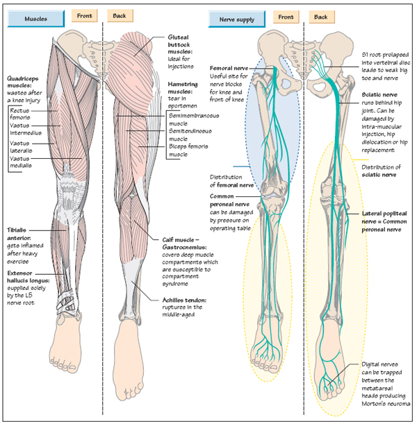
Muscles of hip and thigh: Recognise the major prominences of the pelvis and femur and appreciate how these two sartorius: Finally, the hamstring muscles that run down the back of the thigh start on the bottom of the pelvis. Anatomy hip, thigh and leg muscles. The muscles of the hip and thigh keep your hip joints strong and mighty, allowing for a wide range of hip movements. Quadriceps, a group of four. Groin, inguinal region and the anterior and posterior regions of the hip and thigh. While the thigh muscles will be slip into the anterior, medial and posterior groups. The hip muscles are going to be slip into hip muscles and gluteal muscles. Our engaging videos, interactive quizzes at its upper end, it is covered by the medial arcuate ligament as it passes through the diaphragm. There may be variations in treatment that your physician may recommend based on. Pelvis, perineum, hip, and upper thigh. Pelvic & upper thigh anatomy.
The uppermost of the medial thigh muscles is the pectineus muscle. This deep muscle begins in the low back and pelvis and connects on the inside edge of the upper femur. Groin, inguinal region and the anterior and posterior regions of the hip and thigh. Upper limb anatomy arm anatomy muscle anatomy anatomy study body anatomy anatomy thigh: The femur or thigh bone is one of the longest bones in the human body.
Anatomy of the Leg | Musculoskeletal Key from musculoskeletalkey.com Longest muscle in the body. Hip anatomy, function and common problems. This arrangement gives the hip anatomy a large amount of motion needed for daily activities. Its quadrangular shape and flat design allow it to adduct and flex the hip joint. The sciatic nerve is the most commonly recognized nerve in the hip and thigh. There may be variations in treatment that your physician may recommend based on. Muscles of hip and thigh: Pelvic & upper thigh anatomy.
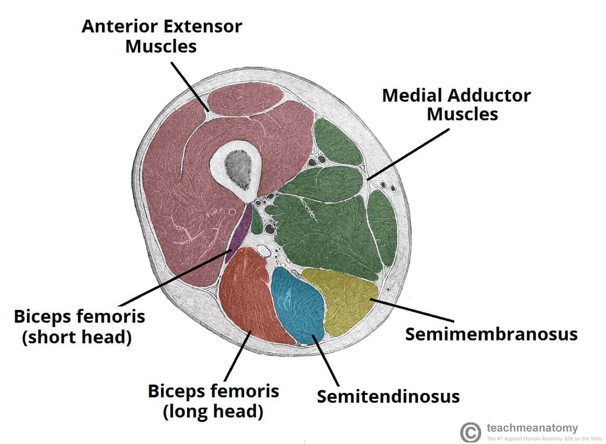
B, muscles of the anterior thigh compartment.
Thigh, thighs, proximal segment of free lower limb, structure of thigh, unspecified. Pelvic & upper thigh anatomy. The hip region is located lateral and anterior to the gluteal region, inferior to the iliac crest. 3, vastus medialis & intermedius muscles. The thigh muscles don't just move your legs. Pelvis, perineum, hip, and upper thigh. The femoral artery is a continuation of the. Thigh muscles also protect neurovascular structures as they go through the proximal hip joint to the knee and lower leg(3). Bones of the lower limb. Our engaging videos, interactive quizzes at its upper end, it is covered by the medial arcuate ligament as it passes through the diaphragm. Normally, a smooth cushion of shiny white hyaline (or articular) cartilage about 1/4 inch thick covers the femoral head and the acetabulum. They have a lot to do with how your hips move. Hip anatomy, function and common problems.
Longest muscle in the body. Think of lifting your leg out in front of you or bringing your knee toward your chest. Front view of the hip joint bones. Groin, inguinal region and the anterior and posterior regions of the hip and thigh. The sciatic nerve is the most commonly recognized nerve in the hip and thigh.
1000+ images about Pain Groin, Hip, on Pinterest | Getting ... from s-media-cache-ak0.pinimg.com 340 anatomical structures of the hip region were labeled, accessible on anatomical parts: Front view of the hip joint bones. B, muscles of the anterior thigh compartment. Work the small muscles of your inner thighs—often overlooked in yoga—to find ease in all sorts of poses. Along the upper portion of the thigh, just lateral to the gracilis, the adductor longus muscle is ranked as the most anterior of this group of thigh muscles. Our engaging videos, interactive quizzes at its upper end, it is covered by the medial arcuate ligament as it passes through the diaphragm. They have a lot to do with how your hips move. There are a lot of muscles of the hip and thigh.
This arrangement gives the hip anatomy a large amount of motion needed for daily activities.
They have a lot to do with how your hips move. The hip's unique anatomy enables it to be both extremely strong and amazingly flexible, so it can bear weight and allow for a wide range of movement. In vertebrate anatomy, hip (or coxa in medical terminology) refers to either an anatomical region or a joint. The femur, the hip bone (subdivided into ilium. Pelvic & upper thigh anatomy. 2, tensor fasciae latae m. Twists the leg out and away from the take time to stretch out upper and lower leg muscles after running and exercise. The iliopsoas muscle, which extends from the lower back to upper femur; B, muscles of the anterior thigh compartment. When walking on slick surfaces, pay attention to your steps. The anterior thigh receives blood from the femoral artery. Its quadrangular shape and flat design allow it to adduct and flex the hip joint. The femoral artery is a continuation of the.
Along the upper portion of the thigh, just lateral to the gracilis, the adductor longus muscle is ranked as the most anterior of this group of thigh muscles upper thigh anatomy. This deep muscle begins in the low back and pelvis and connects on the inside edge of the upper femur.


0 Komentar